Non-Hodgkin’s lymphoma classification using 3D radiomics machine learning models for precision imaging in oncology
BMC Medical Imaging 2025
Abstract
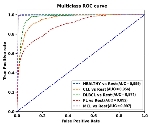
Purpose: To apply quantitative imaging analysis for noninvasive classification of the most frequent subtypes of Non-Hodgkin Lymphoma (NHL) as a basis for a clinical imaging genomic model to support therapeutic monitoring and clinical decision making. Materials and methods: In this single-center study, 201 treatment-naïve patients with biopsy-proven NHL (50 diffuse large B-cell lymphoma [DLBCL], 51 mantle cell lymphoma [MCL], 49 follicular lymphoma [FL], and 51 chronic lymphocytic leukemia [CLL]) and 39 treatment-naïve non-small cell lung cancer patients with positron emission tomography (PET)/computed tomography (CT)-confirmed healthy axillary lymph nodes (LNs) were retrospectively analyzed. Three-dimensional (3D) segmentation and radiomic analysis of pathologically enlarged nodes (n = 1,628) were performed on contrast-enhanced CT scans, including healthy LNs as references. Feature selection was performed using a random forest (RF) classifier. Multiclass Classifier was performed using a Light Gradient Boosting Machine (LGBM) classifier for lymphoma subtype classification. Results: Performance to classify lymphoma from non-lymphoma and lymphoma subtypes was as follows: lymphoma vs. non-lymphoma: area under the curve (AUC) = 0.999; MCL vs. other NHL: AUC = 0.997; DLBCL vs. other NHL: AUC = 0.971; CLL vs. other NHL: AUC = 0.956; FL vs. other NHL: AUC = 0.892. Conclusion : Radiomics combined with multiclass machine learning enables highly accurate, non-invasive differentiation of the major NHL subtypes on routine contrast-enhanced CT. By reliably separating indolent from aggressive phenotypes, this approach lays the groundwork for imaging-genomic models that could streamline biopsy guidance, enhance therapeutic monitoring, and advance precision oncology in lymphoma care.Conducted as a single-centre, retrospective proof-of-concept with internal patient-level cross-validation, these results are promising and form the basis for a prospective multicentre study to confirm generalisability and clinical utility. Clinical relevance statement: Accurate lymphoma classification is essential for predicting clinical behavior and guiding treatment. Imaging aids in disease staging, with quantitative analysis showing promise in predicting pathology and outcome. We explored machine learning on imaging features for lymphoma classification, thus enhancing clinical decisions.
BibTeX
@article{lisson2024non-hodgkin’s,
	title={Non-Hodgkin’s lymphoma classification using 3D radiomics machine learning models for precision imaging in oncology},
	author={Lisson, Christoph Gerhard and G{\"o}tz, Michael and Wolf, Daniel and Manoj, Sabitha and Gallee, Luisa and Andreas Schmidt, Stefan and Tausch, Eugen and Stilgenbauer, Stephan and J. Beer, Ambros and Beer, Meinrad and Sollmann, Nico and Lisson, Cathrina Silvia},
	year={2025},
	journal={BMC Medical Imaging},
	doi={https://doi.org/10.1186/s12880-025-02006-3}
}