CT Radiomics and Clinical Feature Model to Predict Lymph Node Metastases in Early-Stage Testicular Cancer
Onco 2023
Abstract
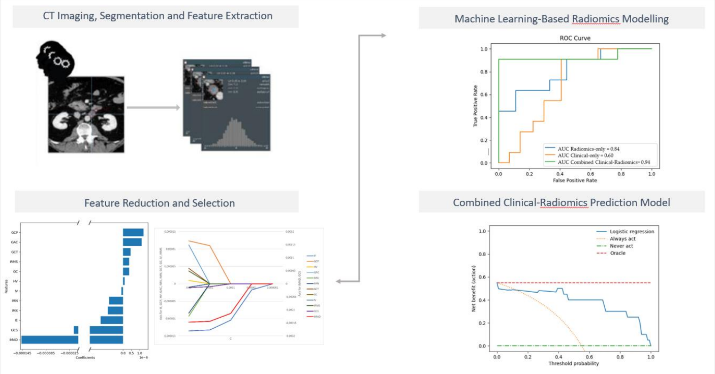
Accurate retroperitoneal lymph node metastasis (LNM) prediction in early-stage testicular germ cell tumours (TGCT) harbours the potential to significantly reduce over- or undertreatment and treatment-related morbidity in this group of young patients as an important survivorship imperative. We investigated the role of computed tomography (CT) radiomics models integrating clinical predictors for the individualized prediction of LNM in early-stage TGCT. Ninety-one patients with surgically proven testicular germ cell tumours and contrast-enhanced CT were included in this retrospective study. Dedicated radiomics software was used to segment 273 retroperitoneal lymph nodes and extract features. After feature selection, radiomics-based machine learning models were developed to predict LN metastasis. The robustness of the procedure was controlled by 10-fold cross-validation. Using multivariable logistic regression modelling, we developed three prediction models: A radiomics-only model, a clinical-only model and a combined radiomics-clinical model. The models’ performance was evaluated using the area under the receiver operating characteristic curve (AUC). Finally, decision curve analysis was performed to estimate the clinical usefulness of the predictive model. The radiomics-only model for predicting lymph node metastasis reached a greater discrimination power than the clinical-only model, with an AUC of 0.84 (± 0.17; 95% CI ) vs 0.60 (± 0.22; 95% CI) in our study cohort. The combined model integrating clinical risk factors and selected radiomics features outperformed the clinical-only and the radiomics-only prediction models and showed good discrimination with an area under the curve of 0.94 ( ± 0.10; 95% CI). The decision curve analysis demonstrated the clinical usefulness of our proposed combined model. The presented combined CT-based radiomics-clinical model represents an exciting non-invasive prediction tool for individualized prediction of LN metastasis in testicular germ cell tumours. Multi-centre validation is required to generate high-quality evidence for its clinical application.
BibTeX
@article{lisson2020ct,
	title={CT Radiomics and Clinical Feature Model to Predict Lymph Node Metastases in Early-Stage Testicular Cancer},
	author={Lisson, Cathrina Silvia and Manoj, Sabitha and Wolf, Daniel and Schrader, Jasper and Andreas Schmidt, Stefan and Beer, Meinrad and G{\"o}tz, Michael and Zengerling, Friedemann and Lisson, Christoph Gerhard},
	year={2023},
	journal={Onco},
	doi={https://doi.org/10.3390/onco3020006}
}