Real-Time Visualization of 3D Amyloid-Beta Fibrils from 2D Cryo-EM Density Maps
Eurographics Workshop on Visual Computing for Biology and Medicine 2020
Abstract
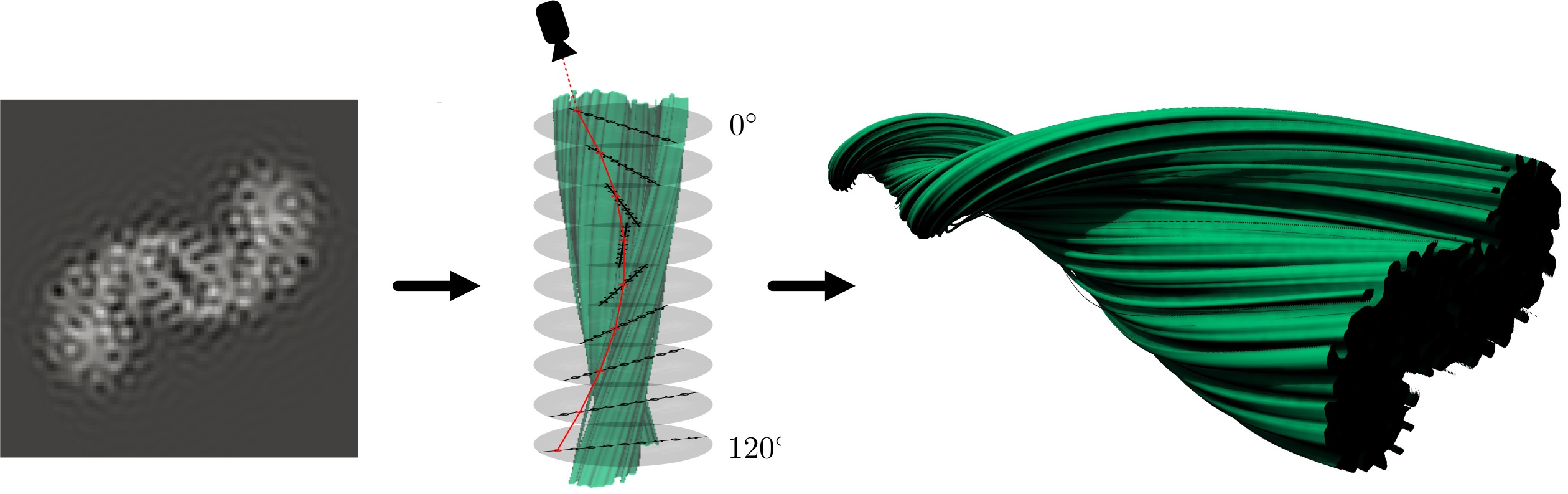
Amyloid-beta fibrils are the result of the accumulation of misfolded amyloid precursor proteins along an axis. These fibrils play a crucial role in the development of Alzheimer's disease, and yet its creation and structure are not fully understood. Visualization is often used to understand the structure of such fibrils. Unfortunately, existing algorithms require high memory consumption limiting their applications. In this paper, we introduce a ray marching algorithm that takes advantage of the inherent repetition in these atomic structures, requiring only a 2D density map to represent the fibril. During ray marching, the texture coordinates are transformed based on the position of the sample along the longitudinal axis, simulating the rotation of the fibrils. Our algorithm reduces memory consumption by a large margin and improves GPU cache hits, making it suitable for real-time visualizations. Moreover, we present several shading algorithms for this type of data, such as shadows or ambient occlusion, in order to improve perception. Lastly, we provide a simple yet effective algorithm to communicate the uncertainty introduced during reconstruction. During the evaluation process, we were able to show, that our approach not only outperforms the Standard Volume Rendering method by significantly lower memory consumption and high image quality for low resolution 2D density maps but also in performance.
BibTeX
@inproceedings{kniesel2020real-time,
	title={Real-Time Visualization of 3D Amyloid-Beta Fibrils from 2D Cryo-EM Density Maps},
	author={Kniesel, Hannah and Ropinski, Timo and Hermosilla, Pedro},
	year={2020},
	pages={115--125},
	editor={Kozl{\'i}kov{\'a}, Barbora and Krone, Michael and Smit, Noeska N. and Nieselt, Kay and Georgia Raidou, Renata}
}